Private Dentmetrik Oral and Dental Health Clinic
Prof. Dr. Beynun Akyavas Avenue No:33/AA, 34680 Uskudar/Istanbul, Turkiye.
©2026 Dentmetrik. All rights reserved.
Cyst and tumor surgeries are the procedures for the complete surgical removal of fluid-filled sacs (cysts) or abnormal tissue growths (tumors) that develop insidiously within the soft tissues of the oral cavity (gums, tongue, lips, cheeks) or inside the jawbones.
Contact Us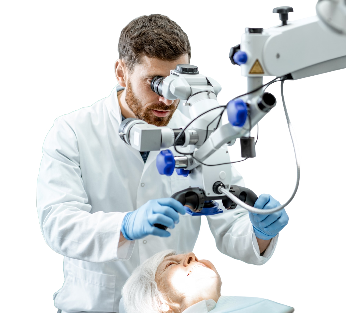

Cyst and tumor surgeries are the procedures for the complete surgical removal of fluid-filled sacs (cysts) or abnormal tissue growths (tumors) that develop insidiously within the soft tissues of the oral cavity (gums, tongue, lips, cheeks) or inside the jawbones. These lesions, which are often detected incidentally during routine dental examinations, can lead to large-scale bone resorption, loss of healthy teeth, and even jaw fractures if left untreated. At Dentmetrik, we utilize advanced 3D radiological imaging (CBCT) systems to diagnose cysts and tumors in the maxillofacial region, and our expert surgeons safely and painlessly remove these pathological formations without damaging surrounding tissues.
Cyst and tumor surgery is the discipline involving the surgical removal (excision or enucleation) of pathological tissues arising in the maxillofacial region due to embryological developmental remnants, trauma, chronic dental infections, or genetic factors. Cysts are benign sacs surrounded by an epithelial membrane and filled with fluid, semi-solid material, or gas. Jaw tumors (which can be benign or malignant) are masses of hard or soft tissue formed by the uncontrolled proliferation of cells. Because the jawbone houses tooth roots, it is anatomically more susceptible to the formation of cysts and tumors compared to other bones in the body.
The primary medical purpose of these critical surgical interventions is to halt the destruction (bone resorption) caused by the pathological formation within the jawbone and to completely eliminate the lesion by delineating its boundaries. Cysts and benign tumors typically grow slowly, but as they expand, they act like a balloon, eroding the surrounding healthy jawbone, putting pressure on nerve canals, and displacing the roots of neighboring teeth. Malignant or aggressive lesions, however, present a much more rapid destructive profile. One of the most important goals of this surgery is to ensure a definitive diagnosis by sending the removed tissue to a pathology laboratory (histopathological examination). Through early surgical cleaning, the integrity of the jawbone is preserved, potential facial asymmetry is prevented, and the cavity created by the lesion is filled with bone grafts (bone powder) to restore the jaw structure to its healthy form.
Cysts and tumors seen in the jaw and oral region are divided into various types based on the tissue of origin (whether they are odontogenic/tooth-related) and their growth characteristics. The primary types of cysts and tumors diagnosed and successfully treated at Dentmetrik clinics include:
Success in cyst and tumor surgery is possible through accurate radiological diagnosis and meticulous surgical planning. The process begins with a detailed clinical examination by our expert surgeons at Dentmetrik. 3D Dental Tomography (CBCT) is an indispensable diagnostic tool for visualizing the exact boundaries of the lesion, the extent of bone resorption, and its relationship with the sinus cavities and the lower jaw nerve (mandibular nerve) in three dimensions. For very large or suspicious-looking lesions, a small piece is taken under local anesthesia (incisional biopsy) and sent to pathology to definitively determine the type of tumor before the main operation.
The scale of the surgical operation depends on the diameter of the lesion. Small and medium-sized cysts are scraped and removed in a single piece along with their membranes (enucleation technique) under local anesthesia in the dental chair, completely painlessly. However, for cysts that occupy a large portion of the jawbone and have reached massive sizes, a special technique called "Marsupialization" may be applied. In this technique, a window is opened into the cyst to drain the fluid, allowing the cyst to shrink on its own over several months; it is then fully removed with a smaller operation. This eliminates the risk of jaw fracture.
Once the cyst or tumor is completely cleared from the jawbone, if the remaining bone cavity is too large for the body to fill on its own, new bone formation is triggered by supporting the space with biocompatible bone grafts and membranes. All removed tissues are sent to fully equipped laboratories for histopathological examination without exception. The recovery process is similar to a routine impacted tooth extraction. Slight swelling and sensitivity are normal in the first few days. Tissues recover quickly with regular use of antibiotics and painkillers. After the final pathology report is issued, the patient's condition is monitored radiologically at specific intervals to confirm the complete healing of the jawbone.
The greatest and most vital advantage of cyst and tumor surgery is the definitive cessation of the destructive process that progresses silently in the jawbones without showing any external symptoms. If these lesions are not removed in time, the jawbone can become as thin as paper, leading to 'pathological fractures' where the jaw breaks suddenly even under minimal chewing force. Surgical intervention completely eliminates this major risk. Additionally, healthy neighboring teeth whose roots have begun to resorb or shift due to the pressure of the cyst are saved, and the internal dental alignment is preserved.
Particularly in tumoral formations, early diagnosis and surgical excision neutralize the risk of a lesion transforming from a benign character to a malignant (cancerous) structure (malignant transformation). Removing soft tissue lesions (such as fibromas) with lasers or cautery relieves the patient from the discomfort of constantly biting that tissue while speaking or eating. A successful surgical operation followed by bone graft treatment restores the jawbone to its former volume and strength, creating a perfect and solid foundation for future implant or prosthetic treatments in that area.
To receive detailed information about cyst and tumor surgery prices in the jaw and facial region, biopsy processes, our advanced radiological diagnostic capabilities, and the treatment privileges offered by our Ankara Dentmetrik clinic to health tourism patients, or to schedule an appointment, contact us immediately.
The vast majority of cysts seen in the jaw are "benign" infection or developmental source lesions and are not cancer (malignant tumors). However, as they grow over time, they melt your jawbone. Every piece removed is necessarily sent to pathology for a definitive diagnosis to ensure your peace of mind.
No, it does not always have to be extracted. In early-diagnosed cysts, the tooth can be saved by applying an apical resection procedure to the root end of the tooth while the cyst is being cleaned. However, if the cyst has completely melted the root of the tooth or the support of the tooth has been lost, a decision to extract may unavoidably be made.
The spaces left after small-sized cysts close on their own as the body produces new bone cells within months. However, in very large cavities (spaces), the area is filled with biocompatible bone powders (grafts) during surgery to increase the resistance of the jaw.
The duration varies depending on the size of the lesion and its location within the bone. An average-sized localized cyst operation under local anesthesia is generally completed successfully between 45 minutes and 1 hour.
The vast majority of cyst and benign tumor operations are performed by approaching from inside the mouth (through the gums). Thanks to this, no stitches, incision marks, or deformities that will be visible from the outside will occur on your face or skin.

We are proud to offer modern facilities which are continually updated, equipped with the latest medical technologies and practices. This commitment to innovation allows us to provide you with the highest standard of care possible.
Please consult your doctor on when you can resume normal activities, as this varies based on the severity of your surgery. For Aesthetic Surgery, training and sports should be avoided for at least eight weeks post-surgery, or as specifically advised for your case. If you have any concerns, please do not hesitate to contact DentMetrik.

In case of any issues when you are back in your home country, you can reach us using the contact numbers provided. The actions we take in response to complications depend on the specific issue.
Once this issue has been raised, we will usually arrange a follow-up consultation with the patient, either via phone or video call, to speak with the surgeon. In some cases, the surgeon may request additional photos of the complication to assess it more thoroughly. Based on the surgeon's evaluation, the patient may receive updated care instructions, additional medication recommendations, be directed to a relevant healthcare facility, or be invited back to our hospital for further treatment if necessary.
Additionally, we issue complication insurance to offer added protection and advantages for our patients, and we stand firmly behind the care we provide. Reach out to us for more information about this.

For Aesthetic Surgery, swelling can last 6 to 12 months. Some patients experience minimal swelling within the first six months, while for others, it may take up to 18 months.
We use cookies
We use cookies to improve your browsing experience and analyse our traffic. By clicking "Accept All", you consent to our use of cookies. Learn more
Kişisel Verilerin Korunması Kanunu Kapsamında
Hasta Aydınlatma Metni
6698 sayılı Kişisel Verilerin Korunması Kanunu (KVKK) uyarınca, kişisel verileriniz; veri sorumlusu olarak ÇENGELKÖY MAH. PROF. DR. BEYNUN AKYAVAŞ CAD. NO:33/AA ÜSKÜDAR adresinde faaliyet gösteren DENTMETRİK SAĞLIK HİZMETLERİ LİMİTED ŞİRKETİ (Klinik) tarafından aşağıda açıklanan kapsamda işlenmektedir.
Bu aydınlatma metni; Klinik'in hastalarına hizmet sunması ve hizmet sunduğu hastalarına ilişkin bu metinde belirtilen kişisel verilerinin sistemine kaydedilmesi kapsamında hastaların KVKK uyarınca aydınlatılması amacıyla hazırlanmıştır.
Kişisel verileriniz, KVKK'da düzenlendiği üzere aşağıda yer alan temel ilkeler doğrultusunda işlenir:
| Veri Kategorisi | Veri Türleri |
|---|---|
| Kimlik | Ad-soyad, TCKN, uyruk, doğum tarihi, cinsiyet |
| İletişim | GSM, e-posta adresi, adres |
| Özlük | Meslek |
| Müşteri İşlem | Hasta tipi, geliş şekli, tavsiye edilen doktor, anlaşmalı kurum, sağlık turizm acentesi, fatura bilgileri, randevu tarihi ve aldığı hizmet, gelecek randevu, yapılan bildirimler, son ziyareti ve ilgilenen doktor |
| Finansal Bilgiler | Borç/bakiye tutarı, tedavi tutarı, toplam ödeme tutarı ve şekli |
| Sağlık Bilgileri | Hastalık bilgisi, geçirdiği hastalıklar/kalıcı hastalıklar, kullandığı ilaçlar, geçirdiği ameliyatlar, kan grubu, tedavi bilgisi, röntgen, rapor, reçete, dişlerin durumu ve daha önce yapılan tedaviler |
| Diğer Bilgiler | Anne adı, baba adı, anne ve baba GSM ve e-posta adresleri, anne ve baba meslek, diş fırçası ve diş macunu türleri, ağız bakım suyu ve diş ipi markası, hobiler |
Klinik, bu aydınlatma metninde belirtilen verileri hastalarından sözlü olarak, yazılı form doldurulması suretiyle, sunulan hizmet kapsamında veya elektronik ortamlar aracılığıyla kendisine iletilmesi ile KVKK'da belirtilen kişisel veri işleme şartlarına uygun bir şekilde elde etmekte ve aşağıda belirtilen amaçlarla uyumlu olarak işlemektedir.
| Veri Kategorileri | İşleme Amaçları | Hukuki Sebepler |
|---|---|---|
| Kimlik, Müşteri İşlem, Finans | İş faaliyetlerinin yürütülmesi/denetimi, finans ve muhasebe işlerinin yürütülmesi, mal/hizmet satış faaliyetlerinin yürütülmesi, saklama ve arşiv faaliyetlerinin yürütülmesi, sözleşme süreçlerinin yürütülmesi, yönetim faaliyetlerinin yürütülmesi | Sözleşmenin kurulması veya ifasıyla doğrudan ilgili olması; Klinik'in hukuki yükümlülüğünü yerine getirebilmesi için zorunlu olması |
| İletişim | İletişim faaliyetlerinin yürütülmesi, iş faaliyetlerinin yürütülmesi/denetimi, mal/hizmet üretim ve operasyon süreçlerinin yürütülmesi, ürün/hizmetlerin pazarlama süreçlerinin yürütülmesi, sözleşme süreçlerinin yürütülmesi | Sözleşmenin kurulması veya ifasıyla doğrudan ilgili olması kaydıyla, sözleşmenin taraflarına ait kişisel verilerin işlenmesinin gerekli olması |
| Özlük, Diğer Bilgiler | İş faaliyetlerinin yürütülmesi/denetimi, müşteri ilişkileri yönetimi süreçlerinin yürütülmesi, müşteri memnuniyetine yönelik aktivitelerin yürütülmesi, saklama ve arşiv faaliyetlerinin yürütülmesi | İlgili kişinin temel hak ve özgürlüklerine zarar vermemek kaydıyla, Klinik'in meşru menfaatleri için veri işlenmesinin zorunlu olması |
| Sağlık Bilgileri | İş faaliyetlerinin yürütülmesi/denetimi, iş sürekliliğinin sağlanması, faaliyetlerin mevzuata uygun yürütülmesi, mal/hizmet satış faaliyetlerinin yürütülmesi, saklama ve arşiv faaliyetlerinin yürütülmesi | Sözleşmenin kurulması veya ifasıyla doğrudan ilgili olması; sizlerden aldığımız açık rızanız |
Klinik, kişisel verilerinizi "bilme gereği" ve "kullanma gereği" ilkelerine uygun olarak, gerekli veri minimizasyonunu sağlayarak ve gerekli teknik ve idari güvenlik tedbirlerini alarak işlemeye özen göstermektedir.
Kişisel verileriniz işbu metinde yer alan işlenme amaçlarının yerine getirilmesi amacıyla yurtiçi veya açık rızanızın alınması kaydıyla yurt dışında bulunan aşağıdaki gruplara aktarılabilir:
Kişisel verilerin korunmasına ilişkin mevzuat kapsamında:
Haklarınıza ilişkin taleplerinizi belirtmek ve kişisel verileriniz üzerindeki haklarınızı kullanmak amacıyla; ÇENGELKÖY MAH. PROF. DR. BEYNUN AKYAVAŞ CAD. A BLOK NO:33/AA ÜSKÜDAR adresine fiziken yazılı olarak, güvenli elektronik imzayla veya mobil imzayla imzalanarak, KEP adresiniz vasıtasıyla veya daha önce bildirilen ve sistemlerimizde kayıtlı bulunan elektronik posta adresinizi kullanmak suretiyle [email protected] adresine e-posta göndererek başvurabilirsiniz.
Klinik, talebinizin niteliğine göre talebi en kısa sürede ve en geç otuz gün içinde ücretsiz olarak sonuçlandıracaktır. Ancak, işlemin ayrıca bir maliyeti gerektirmesi hâlinde, Kişisel Verileri Koruma Kurulu'nca belirlenen tarifedeki ücret alınacaktır.
Açık Rıza Metni: Klinik tarafından "Klinik Hasta Aydınlatma Metni" içerisinde detaylandırılan sağlık bilgilerimin; başta iş faaliyetlerinin yürütülmesi/denetimi, iş sürekliliğinin sağlanması, faaliyetlerin mevzuata uygun yürütülmesi ve mal/hizmet satış faaliyetlerinin yürütülmesi amaçları olmak üzere Klinik Hasta Aydınlatma Metni'nde yer alan kapsam, sınır ve amaçlar doğrultusunda işlenmesine ve aktarılmasına rıza gösteriyorum.
In accordance with the Law No. 6698 on the Protection of Personal Data (KVKK), your personal data is processed by DENTMETRİK SAĞLIK HİZMETLERİ LİMİTED ŞİRKETİ (the Clinic), operating at ÇENGELKÖY MAH. PROF. DR. BEYNUN AKYAVAŞ CAD. NO:33/AA ÜSKÜDAR, as the data controller within the scope explained below.
This information notice has been prepared to inform patients in accordance with KVKK within the scope of the Clinic's provision of services to its patients and the recording of personal data specified in this notice into its systems.
Your personal data is processed in accordance with the following fundamental principles as regulated in KVKK:
| Data Category | Data Types |
|---|---|
| Identity | Full name, national ID number, nationality, date of birth, gender |
| Contact | Mobile phone, email address, address |
| Professional | Occupation |
| Customer Transaction | Patient type, visit type, recommended doctor, affiliated institution, health tourism agency, invoice details, appointment date and received service, future appointments, notifications sent, last visit and attending doctor |
| Financial Information | Debt/balance amount, treatment amount, total payment amount and method |
| Health Information | Disease information, past/chronic diseases, medications used, past surgeries, blood type, treatment information, X-rays, reports, prescriptions, dental condition and previous treatments |
| Other Information | Mother's name, father's name, parents' mobile and email, parents' occupation, toothbrush and toothpaste types, mouthwash and dental floss brand, hobbies |
The Clinic obtains the data specified in this information notice from its patients verbally, through written form completion, within the scope of services provided, or through electronic means, in compliance with the personal data processing conditions specified in KVKK, and processes them in accordance with the purposes stated below.
| Data Categories | Processing Purposes | Legal Grounds |
|---|---|---|
| Identity, Customer Transaction, Financial | Execution/supervision of business activities, finance and accounting operations, sales activities for goods/services, storage and archiving activities, contract processes, management activities | Being directly related to the establishment or performance of a contract; being mandatory for the Clinic to fulfill its legal obligations |
| Contact | Communication activities, execution/supervision of business activities, production and operation processes for goods/services, marketing processes for products/services, contract processes | Processing of personal data of the parties to a contract being necessary, provided it is directly related to the establishment or performance of the contract |
| Professional, Other Information | Execution/supervision of business activities, customer relationship management processes, customer satisfaction activities, storage and archiving activities | Processing being mandatory for the legitimate interests of the Clinic, provided it does not harm the fundamental rights and freedoms of the data subject |
| Health Information | Execution/supervision of business activities, ensuring business continuity, conducting activities in compliance with legislation, sales activities for goods/services, storage and archiving activities | Being directly related to the establishment or performance of a contract; your explicit consent obtained from you |
The Clinic pays attention to processing your personal data in accordance with the principles of "need to know" and "need to use", ensuring the necessary data minimization and taking the necessary technical and administrative security measures.
Your personal data may be transferred to the following groups located domestically or, subject to obtaining your explicit consent, abroad, for the fulfillment of the processing purposes set out in this notice:
Within the scope of legislation on the protection of personal data:
To exercise your rights regarding your personal data, you may apply in writing to ÇENGELKÖY MAH. PROF. DR. BEYNUN AKYAVAŞ CAD. A BLOK NO:33/AA ÜSKÜDAR, or by sending an email signed with a secure electronic signature or mobile signature via your registered email address to [email protected].
The Clinic will conclude your request free of charge as soon as possible and within thirty days at the latest, depending on the nature of the request. However, if the transaction requires an additional cost, the fee determined by the Personal Data Protection Board will be charged.
Explicit Consent Statement: I consent to the processing and transfer of my health information detailed in the "Clinic Patient Information Notice" by the Clinic, in accordance with the scope, limits, and purposes set out in the Clinic Patient Information Notice, primarily for the purposes of executing/supervising business activities, ensuring business continuity, conducting activities in compliance with legislation, and executing sales activities for goods/services.
Gemäß dem Gesetz Nr. 6698 zum Schutz personenbezogener Daten (KVKK) werden Ihre personenbezogenen Daten von der DENTMETRİK SAĞLIK HİZMETLERİ LİMİTED ŞİRKETİ (Klinik), ansässig unter der Adresse ÇENGELKÖY MAH. PROF. DR. BEYNUN AKYAVAŞ CAD. NR:33/AA ÜSKÜDAR, als Verantwortlicher im nachfolgend erläuterten Umfang verarbeitet.
Dieser Informationstext wurde erstellt, um Patienten gemäß KVKK im Rahmen der Leistungserbringung der Klinik und der Erfassung der in diesem Text genannten personenbezogenen Daten in deren Systeme zu informieren.
Ihre personenbezogenen Daten werden gemäß den folgenden im KVKK geregelten Grundprinzipien verarbeitet:
| Datenkategorie | Datenarten |
|---|---|
| Identität | Vollständiger Name, Personalausweisnummer, Staatsangehörigkeit, Geburtsdatum, Geschlecht |
| Kontakt | Mobiltelefon, E-Mail-Adresse, Anschrift |
| Beruf | Beruf |
| Kundentransaktionen | Patiententyp, Besuchsart, empfohlener Arzt, Partnerinstitution, Gesundheitstourismusagentur, Rechnungsdaten, Termindate und erhaltene Leistung, zukünftige Termine, gesendete Benachrichtigungen, letzter Besuch und behandelnder Arzt |
| Finanzinformationen | Schulden-/Saldobetrag, Behandlungsbetrag, Gesamtzahlungsbetrag und -methode |
| Gesundheitsinformationen | Krankheitsinformationen, vergangene/chronische Erkrankungen, verwendete Medikamente, vergangene Operationen, Blutgruppe, Behandlungsinformationen, Röntgenaufnahmen, Berichte, Rezepte, Zahnzustand und frühere Behandlungen |
| Sonstige Informationen | Name der Mutter, Name des Vaters, Mobilnummern und E-Mail-Adressen der Eltern, Beruf der Eltern, Zahnbürsten- und Zahnpastatypen, Mundwasser- und Zahnseide-Marke, Hobbys |
Die Klinik erhebt die in diesem Informationstext genannten Daten von ihren Patienten mündlich, durch Ausfüllen schriftlicher Formulare, im Rahmen der erbrachten Leistungen oder über elektronische Medien, in Übereinstimmung mit den im KVKK festgelegten Bedingungen für die Verarbeitung personenbezogener Daten, und verarbeitet sie gemäß den nachfolgend genannten Zwecken.
| Datenkategorien | Verarbeitungszwecke | Rechtsgrundlagen |
|---|---|---|
| Identität, Kundentransaktionen, Finanzen | Durchführung/Überwachung der Geschäftstätigkeit, Finanz- und Buchhaltungsvorgänge, Verkaufsaktivitäten für Waren/Dienstleistungen, Speicherung und Archivierung, Vertragsprozesse, Managementaktivitäten | Direkter Zusammenhang mit der Begründung oder Erfüllung eines Vertrags; Erforderlichkeit für die Erfüllung der gesetzlichen Verpflichtungen der Klinik |
| Kontakt | Kommunikationsaktivitäten, Durchführung/Überwachung der Geschäftstätigkeit, Produktions- und Betriebsprozesse für Waren/Dienstleistungen, Marketingprozesse für Produkte/Dienstleistungen, Vertragsprozesse | Erforderlichkeit der Verarbeitung personenbezogener Daten der Vertragsparteien, sofern dies in direktem Zusammenhang mit der Begründung oder Erfüllung des Vertrags steht |
| Beruf, Sonstige Informationen | Durchführung/Überwachung der Geschäftstätigkeit, Kundenbeziehungsmanagement, Kundenzufriedenheitsaktivitäten, Speicherung und Archivierung | Erforderlichkeit der Verarbeitung für die berechtigten Interessen der Klinik, sofern die Grundrechte und Grundfreiheiten der betroffenen Person nicht beeinträchtigt werden |
| Gesundheitsinformationen | Durchführung/Überwachung der Geschäftstätigkeit, Sicherstellung der Geschäftskontinuität, gesetzeskonforme Durchführung der Aktivitäten, Verkaufsaktivitäten für Waren/Dienstleistungen, Speicherung und Archivierung | Direkter Zusammenhang mit der Begründung oder Erfüllung eines Vertrags; Ihre von Ihnen erteilte ausdrückliche Einwilligung |
Die Klinik achtet darauf, Ihre personenbezogenen Daten nach den Grundsätzen der „Erforderlichkeit der Kenntnis" und „Erforderlichkeit der Nutzung" zu verarbeiten, die notwendige Datenminimierung sicherzustellen und die erforderlichen technischen und administrativen Sicherheitsmaßnahmen zu treffen.
Ihre personenbezogenen Daten können zur Erfüllung der in diesem Text genannten Verarbeitungszwecke an folgende Gruppen im Inland oder – vorbehaltlich Ihrer ausdrücklichen Einwilligung – im Ausland übermittelt werden:
Im Rahmen der Gesetzgebung zum Schutz personenbezogener Daten:
Um Ihre Rechte in Bezug auf Ihre personenbezogenen Daten auszuüben, können Sie schriftlich an die Adresse ÇENGELKÖY MAH. PROF. DR. BEYNUN AKYAVAŞ CAD. A BLOK NR:33/AA ÜSKÜDAR oder per E-Mail mit sicherer elektronischer Signatur oder mobiler Signatur über Ihre registrierte E-Mail-Adresse an [email protected] einen Antrag stellen.
Die Klinik wird Ihren Antrag je nach Art so schnell wie möglich und spätestens innerhalb von dreißig Tagen kostenlos bearbeiten. Sollte der Vorgang jedoch zusätzliche Kosten verursachen, wird die vom Datenschutzrat festgelegte Gebühr erhoben.
Einwilligungserklärung: Ich willige ein, dass meine im „Patienten-Informationstext der Klinik" aufgeführten Gesundheitsinformationen von der Klinik im Einklang mit dem Umfang, den Grenzen und Zwecken des Patienten-Informationstextes verarbeitet und übermittelt werden, insbesondere zum Zweck der Durchführung/Überwachung der Geschäftstätigkeit, Sicherstellung der Geschäftskontinuität, gesetzeskonformen Durchführung der Aktivitäten und Verkaufsaktivitäten für Waren/Dienstleistungen.
Son güncelleme: 16 Ocak 2025
DentMetrik ("biz", "bize" veya "bizim") https://dentmetrik.com/ web sitesinde ("Hizmet") çerezler kullanmaktadır. Bu siteyi kullanarak çerezlerin kullanımına onay vermiş olursunuz. Bu Çerez Politikası; çerezlerin ne olduğunu, çerezleri nasıl kullandığımızı, iş ortağı olabileceğimiz üçüncü tarafların Hizmet üzerinde çerezleri nasıl kullanabileceğini, çerezlere ilişkin tercihlerinizi ve çerezler hakkında daha fazla bilgiyi açıklamaktadır.
Zorunlu çerezler, güvenli oturum açma veya onay tercihlerinizi ayarlama gibi bu sitenin temel özelliklerini etkinleştirmek için gereklidir. Bu çerezler kişisel olarak tanımlanabilir herhangi bir veri saklamaz.
| Çerez | Süre | Açıklama |
|---|---|---|
| crisp-client* | 6 ay | Crisp, web sitesindeki sohbet işlevi için bu çerezi ayarlar. |
| cookieyes-consent | 1 yıl | CookieYes, kullanıcıların onay tercihlerini hatırlamak için bu çerezi ayarlar; böylece sonraki ziyaretlerde tercihlerine uyulur. Site ziyaretçileri hakkında herhangi bir kişisel bilgi toplamaz veya saklamaz. |
Analiz çerezleri, ziyaretçilerin web sitesiyle nasıl etkileşimde bulunduğunu anlamak için kullanılır. Bu çerezler; ziyaretçi sayısı, hemen çıkma oranı, trafik kaynağı gibi metriklere ilişkin bilgi sağlar.
| Çerez | Süre | Açıklama |
|---|---|---|
| _ga | 1 yıl 1 ay 4 gün | Google Analytics, ziyaretçi, oturum ve kampanya verilerini hesaplamak ve sitenin analiz raporu için site kullanımını izlemek amacıyla bu çerezi ayarlar. Çerez bilgileri anonim olarak saklar ve benzersiz ziyaretçileri tanımak için rastgele oluşturulmuş bir numara atar. |
| _ga_* | 3 ay | Google Tag Manager, hizmetlerini kullanan web sitelerinin reklam verimliliğini denemek amacıyla bu çerezi ayarlar. |
| _ga_* | 1 yıl 1 ay 4 gün | Google Analytics, sayfa görüntülemelerini saklamak ve saymak için bu çerezi ayarlar. |
| _fbp | 3 ay | Facebook, web sitesini ziyaret ettikten sonra Facebook'ta veya Facebook reklamcılığı tarafından desteklenen bir dijital platformda reklam göstermek için bu çerezi ayarlar. |
Reklam çerezleri, daha önce ziyaret ettiğiniz sayfalara göre size özelleştirilmiş reklamlar sunmak ve reklam kampanyalarının etkinliğini analiz etmek için kullanılır.
| Çerez | Süre | Açıklama |
|---|---|---|
| test_cookie | 15 dakika | doubleclick.net, kullanıcının tarayıcısının çerezleri destekleyip desteklemediğini belirlemek için bu çerezi ayarlar. |
| IDE | 1 yıl 24 gün | Google DoubleClick IDE çerezleri, kullanıcının web sitesini nasıl kullandığına dair bilgi depolar ve kullanıcı profiline göre ilgili reklamları sunar. |
DentMetrik ("us", "we", or "our") uses cookies on the https://dentmetrik.com/ website (the "Service"). By using this site, you consent to the use of cookies. This Cookies Policy explains what cookies are, how we use cookies, how third-parties we may partner with may use cookies on the Service, your choices regarding cookies and further information about cookies.
Necessary cookies are required to enable the basic features of this site, such as providing secure log-in or adjusting your consent preferences. These cookies do not store any personally identifiable data.
| Cookie | Duration | Description |
|---|---|---|
| crisp-client* | 6 months | Crisp sets this cookie for chat on website. |
| cookieyes-consent | 1 year | CookieYes sets this cookie to remember users' consent preferences so that their preferences are respected on subsequent visits to this site. It does not collect or store any personal information about the site visitors. |
Analytics cookies are used to understand how visitors interact with the website. These cookies help provide information on metrics such as the number of visitors, bounce rate, traffic source, etc.
| Cookie | Duration | Description |
|---|---|---|
| _ga | 1 year 1 month 4 days | Google Analytics sets this cookie to calculate visitor, session and campaign data and track site usage for the site's analytics report. The cookie stores information anonymously and assigns a randomly generated number to recognize unique visitors. |
| _ga_* | 3 months | Google Tag Manager sets the cookie to experiment advertisement efficiency of websites using their services. |
| _ga_* | 1 year 1 month 4 days | Google Analytics sets this cookie to store and count page views. |
| _fbp | 3 months | Facebook sets this cookie to display advertisements when either on Facebook or on a digital platform powered by Facebook advertising after visiting the website. |
Advertisement cookies are used to provide visitors with customised advertisements based on the pages you visited previously and to analyse the effectiveness of the ad campaigns.
| Cookie | Duration | Description |
|---|---|---|
| test_cookie | 15 minutes | doubleclick.net sets this cookie to determine if the user's browser supports cookies. |
| IDE | 1 year 24 days | Google DoubleClick IDE cookies store information about how the user uses the website to present them with relevant ads according to the user profile. |
DentMetrik („uns", „wir" oder „unser") verwendet Cookies auf der Website https://dentmetrik.com/ (der „Dienst"). Durch die Nutzung dieser Website stimmen Sie der Verwendung von Cookies zu. Diese Cookie-Richtlinie erklärt, was Cookies sind, wie wir Cookies verwenden, wie Drittanbieter, mit denen wir zusammenarbeiten, Cookies im Dienst verwenden können, Ihre Wahlmöglichkeiten bezüglich Cookies und weitere Informationen über Cookies.
Notwendige Cookies sind erforderlich, um die grundlegenden Funktionen dieser Website zu ermöglichen, wie z. B. die sichere Anmeldung oder die Anpassung Ihrer Einwilligungseinstellungen. Diese Cookies speichern keine personenbezogenen Daten.
| Cookie | Dauer | Beschreibung |
|---|---|---|
| crisp-client* | 6 Monate | Crisp setzt dieses Cookie für die Chat-Funktion auf der Website. |
| cookieyes-consent | 1 Jahr | CookieYes setzt dieses Cookie, um die Einwilligungseinstellungen der Benutzer zu speichern, damit ihre Präferenzen bei nachfolgenden Besuchen berücksichtigt werden. Es werden keine persönlichen Daten der Website-Besucher erfasst oder gespeichert. |
Analyse-Cookies werden verwendet, um zu verstehen, wie Besucher mit der Website interagieren. Diese Cookies liefern Informationen zu Metriken wie Besucherzahl, Absprungrate, Verkehrsquelle usw.
| Cookie | Dauer | Beschreibung |
|---|---|---|
| _ga | 1 Jahr 1 Monat 4 Tage | Google Analytics setzt dieses Cookie, um Besucher-, Sitzungs- und Kampagnendaten zu berechnen und die Website-Nutzung für den Analysebericht zu verfolgen. Das Cookie speichert Informationen anonym und weist eine zufällig generierte Nummer zu, um eindeutige Besucher zu erkennen. |
| _ga_* | 3 Monate | Google Tag Manager setzt dieses Cookie, um die Werbeeffizienz von Websites zu testen, die ihre Dienste nutzen. |
| _ga_* | 1 Jahr 1 Monat 4 Tage | Google Analytics setzt dieses Cookie, um Seitenaufrufe zu speichern und zu zählen. |
| _fbp | 3 Monate | Facebook setzt dieses Cookie, um nach dem Besuch der Website Werbung auf Facebook oder auf einer von Facebook-Werbung betriebenen digitalen Plattform anzuzeigen. |
Werbe-Cookies werden verwendet, um Besuchern personalisierte Werbung basierend auf zuvor besuchten Seiten anzuzeigen und die Wirksamkeit von Werbekampagnen zu analysieren.
| Cookie | Dauer | Beschreibung |
|---|---|---|
| test_cookie | 15 Minuten | doubleclick.net setzt dieses Cookie, um festzustellen, ob der Browser des Benutzers Cookies unterstützt. |
| IDE | 1 Jahr 24 Tage | Google DoubleClick IDE-Cookies speichern Informationen darüber, wie der Benutzer die Website nutzt, um ihm gemäß seinem Benutzerprofil relevante Werbung anzuzeigen. |
Özel Dentmetrik Ağız ve Diş Sağlığı Polikliniği
Hasta Tedavi Şartları ve Garanti Politikası
İşbu Tedavi Koşulları ve Politikalar metni, ÇENGELKÖY MAH. PROF. DR. BEYNUN AKYAVAŞ CAD. NO:33/AA ÜSKÜDAR adresinde faaliyet gösteren DENTMETRİK SAĞLIK HİZMETLERİ LİMİTED ŞİRKETİ (bundan sonra "Klinik" olarak anılacaktır) tarafından sunulan ağız ve diş sağlığı hizmetlerine ilişkin koşulları, hasta sorumluluklarını, garanti politikasını ve tedavi sürecine dair hak ve yükümlülükleri düzenlemektedir.
Bu metin kapsamında yer alan tedavi hizmetleri şunlardır:
Klinik, tedaviye başlamadan önce hastayı tedavi planı, olası riskler, alternatif yöntemler ve tahmini maliyet hakkında bilgilendirir. Hasta, tedavi planını onayladığına dair yazılı veya elektronik onay verir. Tedaviye başlanması, hastanın bu koşulları kabul ettiği anlamına gelir.
Tedavi süresince ortaya çıkabilecek beklenmedik durumlar (ek tedavi gereksinimi, tedavi planında değişiklik vb.) hastaya derhal bildirilir ve hastanın onayı alınmadan ek işlem yapılmaz.
Tedavinin başarısı büyük ölçüde hasta uyumuna bağlıdır. Hasta aşağıdaki yükümlülükleri kabul eder:
İmplant kaynaklı komplikasyonlarda (implant kaybı, osseointegrasyon başarısızlığı), tedavi tarihinden itibaren 2 yıl içinde başvurulması kaydıyla, aynı bölgeye yeniden implant uygulaması ücretsiz olarak gerçekleştirilir. İmplant üst yapıları (kron, köprü) için garanti süresi 2 yıldır.
Protetik restorasyonlarda malzeme veya teknik kaynaklı sorunlar (kırılma, dökülme, uyumsuzluk) için garanti süresi teslim tarihinden itibaren 2 yıldır. Bu süre içinde ücretsiz onarım veya yeniden yapım sağlanır.
Kanal tedavisi uygulanan dişlerde, tedaviden kaynaklanan bir sorun tespit edilmesi halinde, tedavi tarihinden itibaren 1 yıl içinde başvurulması kaydıyla yeniden tedavi ücretsiz olarak yapılır.
Dolgu tedavilerinde, dolgunun düşmesi veya kırılması durumunda, uygulama tarihinden itibaren 1 yıl içinde başvurulması kaydıyla ücretsiz yenileme yapılır.
Ortodontik tedavilerde, tedavi planına uyulması ve kontrol randevularına düzenli gelinmesi koşuluyla, planlanan tedavi hedefine ulaşılana kadar tedavi sürdürülür. Hastanın randevularına gelmemesi veya aparey kullanım talimatlarına uymaması durumunda tedavi sonuçları garanti kapsamı dışında kalır.
Aşağıdaki durumlarda tedavi garantisi geçerli değildir:
Klinik, randevu sistemiyle çalışmaktadır. Hastalarımızdan aşağıdaki kurallara uyması beklenmektedir:
Tedavi ücretleri, tedavi planının onaylanmasının ardından hastaya bildirilir. Klinik aşağıdaki ödeme yöntemlerini kabul etmektedir:
Uzun süreli tedavilerde aşamalı ödeme planı uygulanabilir. Tedaviye başlanabilmesi için belirlenen başlangıç ödemesinin yapılmış olması gerekmektedir. Tedavi yarıda bırakılması halinde, o tarihe kadar gerçekleştirilen işlemlerin ücreti tam olarak tahsil edilir.
Klinik ve hekimleri, tıbbi standartlara ve güncel diş hekimliği protokollerine uygun olarak tedavi hizmeti sunmaktadır. Her klinik prosedür belirli riskler taşımakta olup, tedavi sonuçları hastanın bireysel yapısına, sağlık durumuna ve tedaviye uyumuna göre farklılık gösterebilir. Belirli bir tedavi sonucu hedef niteliğinde olup garanti edilemez.
Klinik, mücbir sebep halleri (doğal afet, salgın hastalık, yasal düzenleme değişiklikleri vb.) nedeniyle hizmet sunumunda aksaklık yaşanması durumunda sorumlu tutulamaz.
Tedavi sürecinize ilişkin her türlü soru, öneri ve şikayetleriniz için aşağıdaki iletişim kanallarından bize ulaşabilirsiniz:
Adres: Çengelköy Mah. Prof. Dr. Beynun Akyavaş Cad. No:33/AA, 34680 Üsküdar/İstanbul
Telefon: +90 216 766 41 00 / +90 532 376 87 65
E-posta: [email protected]
İşbu Tedavi Koşulları ve Politikalar metni, Klinik tarafından gerekli görüldüğünde güncellenebilir. Güncel metin her zaman Klinik'in internet sitesinde ve fiziki ortamında mevcuttur. Tedaviye başlanması, hastanın bu koşulları okuduğu ve kabul ettiği anlamına gelir.
This Treatment Terms and Policies document regulates the conditions regarding oral and dental health services provided by DENTMETRİK SAĞLIK HİZMETLERİ LİMİTED ŞİRKETİ (hereinafter referred to as the "Clinic"), operating at ÇENGELKÖY MAH. PROF. DR. BEYNUN AKYAVAŞ CAD. NO:33/AA ÜSKÜDAR, including patient responsibilities, warranty policy, and rights and obligations pertaining to the treatment process.
The treatment services covered under this document are:
The Clinic informs the patient about the treatment plan, potential risks, alternative methods, and estimated costs before commencing treatment. The patient provides written or electronic consent confirming their approval of the treatment plan. The commencement of treatment constitutes acceptance of these terms.
Unexpected situations that may arise during treatment (need for additional treatment, changes to the treatment plan, etc.) are immediately communicated to the patient, and no additional procedures are performed without the patient's consent.
The success of treatment depends largely on patient compliance. The patient agrees to the following obligations:
In cases of implant-related complications (implant loss, osseointegration failure), re-implantation in the same area is performed free of charge, provided the patient applies within 2 years of the treatment date. The warranty period for implant superstructures (crowns, bridges) is 2 years.
The warranty period for material or technical issues (fracture, debonding, mismatch) in prosthetic restorations is 2 years from the date of delivery. Free repair or remake is provided within this period.
If a treatment-related issue is identified in teeth that have undergone root canal treatment, retreatment is performed free of charge, provided the patient applies within 1 year of the treatment date.
In cases where a filling falls out or fractures, free replacement is provided, provided the patient applies within 1 year of the application date.
For orthodontic treatments, treatment continues until the planned treatment goals are achieved, provided the treatment plan is followed and follow-up appointments are attended regularly. Treatment outcomes are excluded from warranty coverage if the patient fails to attend appointments or does not comply with appliance usage instructions.
The treatment warranty does not apply in the following situations:
The Clinic operates on an appointment basis. Patients are expected to adhere to the following rules:
Treatment fees are communicated to the patient upon approval of the treatment plan. The Clinic accepts the following payment methods:
Instalment payment plans may be applied for long-term treatments. The designated initial payment must be completed before treatment can commence. In the event that treatment is discontinued, the full fee for procedures performed up to that date will be charged.
The Clinic and its dentists provide treatment services in accordance with medical standards and current dental protocols. Every clinical procedure carries certain risks, and treatment outcomes may vary depending on the patient's individual constitution, health condition, and compliance with treatment. A specific treatment outcome is a target and cannot be guaranteed.
The Clinic cannot be held liable for disruptions in service delivery due to force majeure events (natural disasters, epidemics, changes in legal regulations, etc.).
For any questions, suggestions, or complaints regarding your treatment process, you may reach us through the following contact channels:
Address: Çengelköy Mah. Prof. Dr. Beynun Akyavaş Cad. No:33/AA, 34680 Üsküdar/İstanbul
Phone: +90 216 766 41 00 / +90 532 376 87 65
Email: [email protected]
This Treatment Terms and Policies document may be updated by the Clinic when deemed necessary. The current version is always available on the Clinic's website and at its physical premises. The commencement of treatment constitutes the patient's acknowledgment and acceptance of these terms.
Dieses Dokument zu Behandlungsbedingungen und Richtlinien regelt die Bedingungen für die von der DENTMETRİK SAĞLIK HİZMETLERİ LİMİTED ŞİRKETİ (im Folgenden „Klinik" genannt), ansässig unter ÇENGELKÖY MAH. PROF. DR. BEYNUN AKYAVAŞ CAD. NR:33/AA ÜSKÜDAR, erbrachten Mund- und Zahngesundheitsleistungen, einschließlich der Patientenverantwortlichkeiten, der Garantierichtlinie sowie der Rechte und Pflichten im Zusammenhang mit dem Behandlungsprozess.
Die unter diesem Dokument abgedeckten Behandlungsleistungen sind:
Die Klinik informiert den Patienten vor Behandlungsbeginn über den Behandlungsplan, mögliche Risiken, alternative Methoden und die geschätzten Kosten. Der Patient erteilt seine schriftliche oder elektronische Zustimmung zum Behandlungsplan. Der Behandlungsbeginn gilt als Annahme dieser Bedingungen.
Unerwartete Situationen, die während der Behandlung auftreten können (Notwendigkeit einer Zusatzbehandlung, Änderungen am Behandlungsplan usw.), werden dem Patienten unverzüglich mitgeteilt, und ohne die Zustimmung des Patienten werden keine zusätzlichen Eingriffe vorgenommen.
Der Behandlungserfolg hängt maßgeblich von der Mitwirkung des Patienten ab. Der Patient erklärt sich mit folgenden Pflichten einverstanden:
Bei implantatbedingten Komplikationen (Implantatverlust, Versagen der Osseointegration) wird die erneute Implantation im selben Bereich kostenlos durchgeführt, sofern der Patient sich innerhalb von 2 Jahren nach dem Behandlungsdatum meldet. Die Garantiezeit für Implantat-Suprastrukturen (Kronen, Brücken) beträgt 2 Jahre.
Die Garantiezeit für material- oder technikbedingte Probleme (Bruch, Ablösung, Passungenauigkeit) bei prothetischen Restaurationen beträgt 2 Jahre ab dem Lieferdatum. Innerhalb dieses Zeitraums wird eine kostenlose Reparatur oder Neuanfertigung gewährleistet.
Wird bei wurzelkanalbehandelten Zähnen ein behandlungsbedingtes Problem festgestellt, wird die Nachbehandlung kostenlos durchgeführt, sofern der Patient sich innerhalb von 1 Jahr nach dem Behandlungsdatum meldet.
Bei Herausfallen oder Bruch einer Füllung wird ein kostenloser Ersatz gewährt, sofern der Patient sich innerhalb von 1 Jahr nach dem Anwendungsdatum meldet.
Bei kieferorthopädischen Behandlungen wird die Behandlung bis zum Erreichen der geplanten Behandlungsziele fortgesetzt, sofern der Behandlungsplan eingehalten und die Kontrolltermine regelmäßig wahrgenommen werden. Behandlungsergebnisse sind von der Garantie ausgeschlossen, wenn der Patient Termine versäumt oder die Gebrauchsanweisungen für Apparaturen nicht befolgt.
Die Behandlungsgarantie gilt nicht in folgenden Situationen:
Die Klinik arbeitet auf Terminbasis. Von unseren Patienten wird die Einhaltung folgender Regeln erwartet:
Die Behandlungskosten werden dem Patienten nach Genehmigung des Behandlungsplans mitgeteilt. Die Klinik akzeptiert folgende Zahlungsmethoden:
Für Langzeitbehandlungen können Ratenzahlungspläne angeboten werden. Die festgelegte Anfangszahlung muss vor Behandlungsbeginn geleistet werden. Im Falle eines Behandlungsabbruchs wird die volle Gebühr für bis zu diesem Zeitpunkt durchgeführte Eingriffe berechnet.
Die Klinik und ihre Zahnärzte erbringen Behandlungsleistungen gemäß medizinischen Standards und aktuellen zahnmedizinischen Protokollen. Jeder klinische Eingriff birgt bestimmte Risiken, und die Behandlungsergebnisse können je nach individueller Konstitution, Gesundheitszustand und Behandlungscompliance des Patienten variieren. Ein bestimmtes Behandlungsergebnis ist ein Ziel und kann nicht garantiert werden.
Die Klinik kann nicht für Unterbrechungen der Leistungserbringung aufgrund höherer Gewalt (Naturkatastrophen, Epidemien, Änderungen der Rechtsvorschriften usw.) haftbar gemacht werden.
Für alle Fragen, Anregungen oder Beschwerden zu Ihrem Behandlungsprozess erreichen Sie uns über folgende Kontaktkanäle:
Adresse: Çengelköy Mah. Prof. Dr. Beynun Akyavaş Cad. Nr:33/AA, 34680 Üsküdar/İstanbul
Telefon: +90 216 766 41 00 / +90 532 376 87 65
E-Mail: [email protected]
Dieses Dokument zu Behandlungsbedingungen und Richtlinien kann von der Klinik bei Bedarf aktualisiert werden. Die aktuelle Fassung ist stets auf der Website der Klinik und in deren Räumlichkeiten verfügbar. Der Behandlungsbeginn gilt als Kenntnisnahme und Akzeptanz dieser Bedingungen durch den Patienten.